
Why Ultrasound Training Requires More Than Reading Images

Ultrasound can look deceptively simple from the outside. A clinician places a probe on the patient, an image appears on the screen, and a report is produced afterward. For patients, the most visible part of the process is often the final image or written result. For the person performing the examination, however, ultrasound involves a much broader set of skills.
Being able to recognize an organ or abnormality on a saved image is only one part of sonography. The operator must first find the correct anatomical structure, position and move the probe appropriately, optimize the image, recognize artifacts, and decide whether the views obtained are sufficient for interpretation. These skills develop through practical training and repetition rather than image reading alone.
Reading the Image Is Only One Part of the Process
There is an important difference between interpreting an ultrasound image and acquiring one.
When students look at a textbook image, the difficult work has already been done. Someone has located the structure, chosen an appropriate view, adjusted the machine settings, and captured an image that clearly demonstrates the anatomy or pathology being discussed.
During a real examination, none of those steps happen automatically.
The clinician starts with a patient and a probe. The target structure may not immediately be visible. Its appearance can change according to the angle of the probe, the patient’s position, body characteristics, and the surrounding anatomy.
The operator therefore has to create the useful image before it can be interpreted.
This is one reason ultrasound has a significant hands-on component. A learner who can correctly identify the liver, kidney, or gallbladder on prepared images may still have difficulty locating and displaying those structures during an actual examination.
Training needs to develop both abilities.
Probe Positioning Changes What the Clinician Sees
The ultrasound probe is more than a camera pointed at an organ. Small movements can considerably change the information displayed on the screen.
Operators use several types of movements while scanning. They may slide the probe to a different location, rotate it, tilt it, change its angle, or apply different amounts of pressure.
Each movement affects the ultrasound beam and therefore the resulting image.
For example, a structure that appears poorly defined from one angle may become much clearer after a slight adjustment. An organ may also need to be examined in several planes to understand its shape and relationship with surrounding structures.
Patient positioning matters as well.
A clinician may ask someone to turn onto one side, take a deep breath, or briefly hold their breath. These requests are often made because changing the position of the body or internal organs can create a better acoustic window.
Learning these techniques requires coordination between what the hands are doing and what the eyes are seeing.
A student must gradually learn that a particular movement of the probe produces a predictable change on the screen.
That connection is difficult to develop by studying static images.
Good Ultrasound Training Builds Anatomical Awareness
Ultrasound also requires a strong understanding of anatomy.
Medical students may initially learn anatomy from diagrams, textbooks, cadavers, or three-dimensional models. During sonography, however, anatomy appears very differently.
An ultrasound image shows a relatively small section of the body at a particular angle.
The learner has to understand where that section fits within the larger three-dimensional anatomy of the patient.
This becomes especially important because the same organ can look quite different depending on the imaging plane.
A transverse view may have a completely different appearance from a longitudinal view. Nearby vessels or tissues can also appear similar until the learner understands their location and characteristics.
Experienced operators therefore do more than memorize what individual organs look like.
They develop a mental map of the anatomy and learn how moving the probe changes the section being displayed.
This spatial understanding helps them navigate from one structure to another and recognize when the image does not correspond to the expected anatomy.
Artifacts Can Look Like Real Findings
Another important part of ultrasound education is learning about artifacts.
Ultrasound images are created from sound waves interacting with tissues. Because of the physics involved, the screen may sometimes display features that do not directly correspond to a physical structure.
Artifacts can produce shadows, echoes, apparent duplications, unusual brightness, or distorted borders.
For a beginner, some of these effects may resemble pathology.
An experienced operator learns to recognize common artifacts and may change the probe position or machine settings to determine whether a suspicious feature is real.
This is another reason why interpretation and image acquisition are closely connected.
When clinicians control the scan themselves, they can investigate what they are seeing. They can approach the area from another direction, change the imaging plane, or compare several views.
A single static image does not always provide that opportunity.
Understanding artifacts therefore requires both theoretical knowledge and practical experience.
Real Skill Comes From Repetition
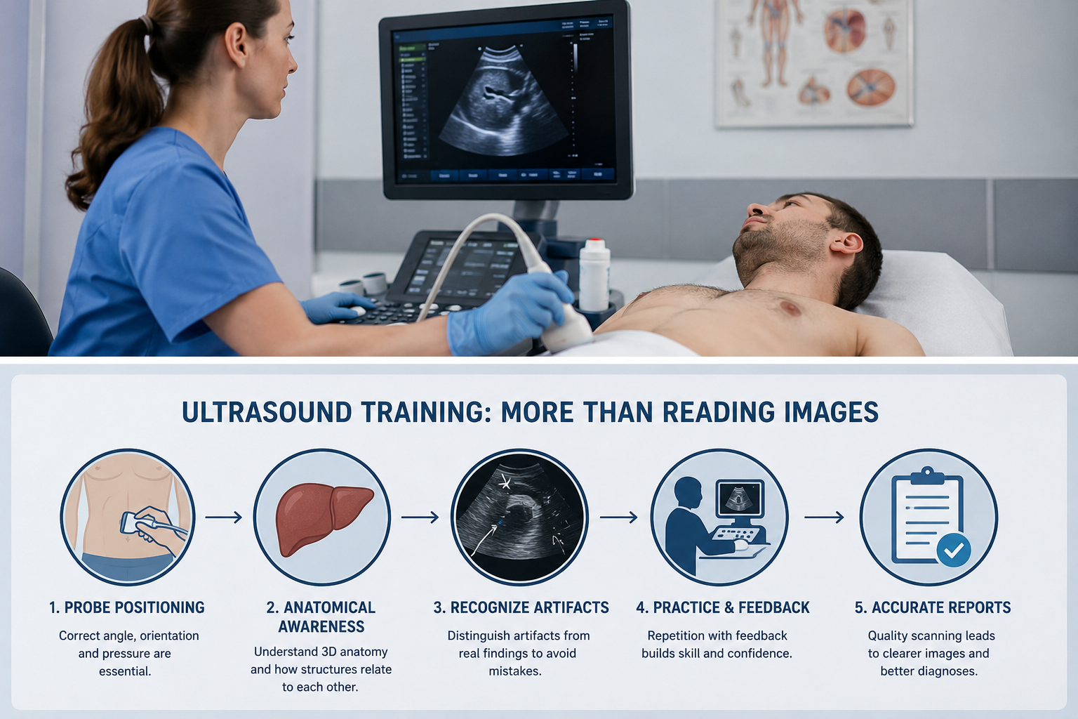
Most practical medical skills improve with repetition, and ultrasound is no exception.
A student needs time to become comfortable holding and controlling the probe while simultaneously watching the screen. At first, even small adjustments can be difficult because the learner is concentrating on several tasks at once.
With practice, those movements become more intuitive.
Repetition can help learners develop skills such as:
- locating anatomical structures efficiently
- maintaining the correct orientation
- controlling probe pressure and movement
- obtaining standard imaging planes
- adjusting the image when visualization is poor
- distinguishing normal anatomy from unusual findings
- recognizing common artifacts
- performing a structured examination without missing important areas
Repeated practice is particularly valuable because learners do not progress at exactly the same speed.
One person may understand image interpretation quickly but struggle with probe control. Another may acquire technically good images but need more time to recognize anatomical landmarks.
Training allows each learner to repeat the areas that need the most improvement.
Simulation Connects Hand Movement With Screen Results
Simulation can be particularly useful during the early stages of ultrasound education because it gives learners an opportunity to practice scanning in a controlled setting.
The important feature is interaction.
Instead of simply being shown an ultrasound image, the learner moves a probe and observes how those movements affect what appears on the screen. This helps establish the connection between anatomy, probe position, and image acquisition.
For example, training with such ultrasound simulator can allow learners to practice probe positioning, image optimization, anatomical identification, and different clinical scenarios without depending on the availability of a particular patient or finding.
SonoVision, for example, includes real-time probe tracking, three-dimensional organ models and multiple clinical scenarios designed around practical ultrasonography training.
Simulation can also make repetition easier.
A difficult view can be attempted several times. The learner can change the probe angle, compare the resulting images, and immediately try again.
The same anatomy can then be revisited during another session.
This does not replace scanning real patients. Human anatomy varies, and communication with patients is an important part of clinical practice.
Instead, simulation can help students build basic technical skills before they apply them in more complex clinical environments.
Training Must Include Image Quality
Getting any image onto the screen is not the same as obtaining a diagnostically useful image.
Operators must learn how to improve what they see.
That can involve adjusting probe position as well as changing imaging parameters. The goal is to display the relevant anatomy clearly enough for appropriate assessment and documentation.
Professional ultrasound guidance reflects this emphasis on examination quality. The American Institute of Ultrasound in Medicine practice parameters provide guidance for the performance and recording of high-quality ultrasound examinations and describe minimum criteria for complete examinations across different applications.
This is an important point for learners because technical quality and interpretation cannot be separated completely.
When the necessary anatomy is poorly visualized, interpretation becomes more difficult.
A trained operator therefore continually evaluates the image during the examination rather than waiting until the scan is finished.
Why This Matters for the Medical Report
Patients usually receive the final result of an ultrasound examination as a written report, sometimes accompanied by selected images.
It can be easy to think that the report is produced simply by looking at those images.
In reality, the quality of the examination begins much earlier.
The person performing the scan has to obtain appropriate views, examine the relevant anatomy, capture useful images, take measurements when required, and document important findings.
The interpreting clinician then works with the information generated during that examination.
Poor visualization does not automatically mean that a report will be wrong, but limited or inadequate images can make confident interpretation more difficult.
This is why practical ultrasound skills matter even to people who are mainly interested in understanding their final medical report.
The report represents the end of a process that started with image acquisition.
Good training helps make that process more consistent.
Ultrasound Is Different From Many Other Imaging Methods
Ultrasound has another characteristic that makes practical training particularly important: the examination develops in real time.
With some other imaging methods, a predefined series of images is acquired and then reviewed.
During ultrasound, the operator is actively searching while the examination is taking place.
What appears on the screen influences the next movement.
The clinician may identify one structure, move to another area, return to the original location, change the angle, make a measurement, or investigate something unexpected.
This makes ultrasound an interactive examination.
The operator is continuously making small decisions about where to look and what to record.
A learner therefore needs to develop a scanning strategy rather than simply recognize individual pictures.
That strategy becomes increasingly important in more complicated examinations where several structures need to be assessed systematically.
What Patients Should Understand About an Ultrasound Examination
Patients do not need to understand every technical aspect of sonography, but knowing how the examination works can make some parts of the experience less confusing.
For example, the clinician may spend considerably longer examining one area than another. That does not necessarily mean something abnormal has been found. The structure may simply require several views.
The operator may also repeatedly return to the same location.
Again, this can be part of obtaining the necessary imaging planes or measurements.
Requests to change position, breathe differently, or remain still can also be related to image quality.
And the quality of the ultrasound machine is only one factor in the examination.
The knowledge and practical skill of the person using it are also important.
For that reason, ultrasound education involves much more than teaching students what various diseases look like on a screen.
Simulation and Clinical Experience Work Together
There is no substitute for eventually working with real patients.
Real examinations introduce anatomical variation, communication challenges, different body types, unexpected findings, and practical situations that cannot be reproduced perfectly in a training environment.
Simulation serves a different purpose.
It gives learners a place to build foundational skills before every mistake or hesitation affects an actual examination.
Students can become familiar with the relationship between their hand movements and the resulting image. They can practice structured scanning sequences and repeat difficult tasks.
Once those skills are more stable, clinical training allows them to apply what they have learned to real people.
The two approaches can therefore complement each other.
Simulation provides controlled repetition, while clinical experience provides real-world variability.
Conclusion
Learning ultrasound involves far more than looking at images and deciding what they show.
Before an image can be interpreted, someone has to create it. That requires knowledge of anatomy, control of the probe, understanding of imaging planes, recognition of artifacts, image optimization, and a systematic approach to the examination.
These abilities are developed through hands-on practice.
Simulation can support that process by allowing learners to repeat scans and observe how changes in probe position affect the image before moving into real clinical practice.
For patients, this also helps explain why an ultrasound report depends on more than the final pictures attached to it. The quality of the examination begins with the way those images are obtained.
Reading ultrasound images is an important skill. Learning how to consistently produce the right images in the first place is equally important.